Trust the Experts in Biomarker Services
Founded to provide excellence in GI biopsy processing for histopathology endpoints,
AcelaBio fully supports tissue analysis for Alimentiv studies.
From reliable images for central reader scoring to samples for advanced exploratory endpoints,
see what AcelaBio’s expertise can do for your program.
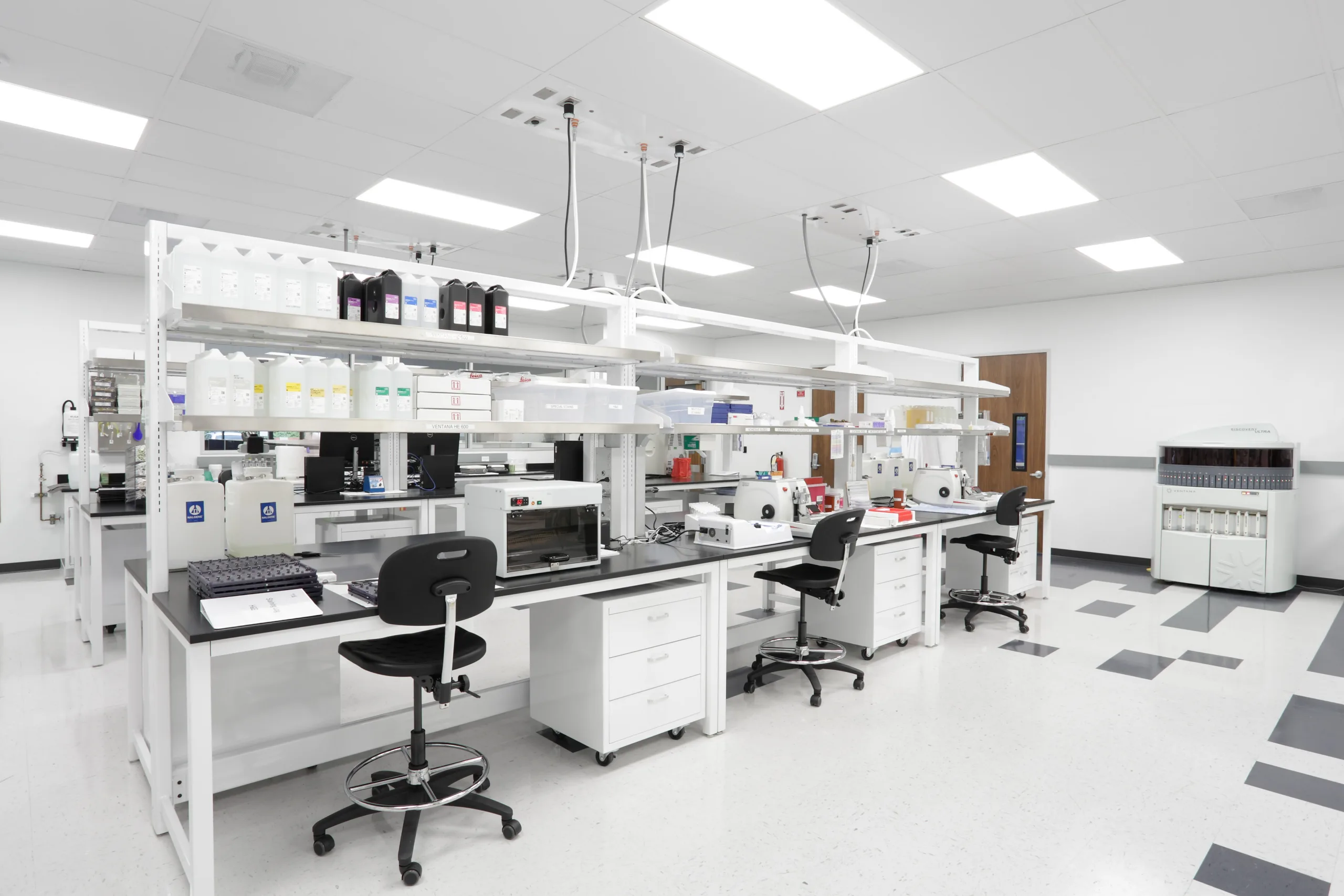
Premier Histopathology Laboratory Services
GI tissues present unique challenges: small tissue size, delicate epithelial structures, and region-specific architecture that require specialized handling. High-quality processing is essential to preserve morphology, minimize artifacts, and ensure that pathological features and biomarker signals remain intact — and AcelaBio delivers.
As a histopathology leader and pioneer, AcelaBio offers high-quality biopsy processing, embedding, sectioning, and staining to generate consistent, publication-grade slides and images for research and clinical development:
Harmonized SOPs and rigorous quality controls tailored to the unique characteristics of GI biopsies ensure reproducibility within and across studies.
A clear chain of custody, following 21 CFR Part 11 guidelines, keeps your blocks, slides, and data audit ready.
Standardized workflows are utilized to upload whole slide images for central reading and scoring utilizing Lucidity™ and Notō, Alimentiv’s proprietary, multimodal imaging platform for capture and review across all modalities.
As a small, specialized laboratory, AcelaBio spots processing challenges early, preventing downstream delays and protecting the integrity of your samples.
AcelaBio’s GI-focused workflows optimize the preservation of RNA, protein, and spatial architecture, enabling downstream assays critical for biomarker discovery, mechanisms-of-action studies, and translational research.
AcelaBio’s services are designed to extract maximum value from each biopsy, enabling histopathology, immunostaining, and spatial biology assays all from the same tissue block with:
- Advanced tissue-based assays to generate richer biological insights without the need for additional sample collection
- On-site analysis to reduce turnaround time, lower per-sample costs, and eliminate risks associated with shipping tissue samples between vendors
- Optimized microtomy and block utilization strategies to ensure efficient use of each tissue block
Immunostaining and Digital Image Analysis
AcelaBio offers a range of immunohistochemistry (IHC), immunofluorescence (IF), in situ hybridization (ISH), and special stains to precisely characterize GI tissues:
Customized solutions
Expert scientists work closely with you to select, optimize, or validate the most appropriate markers for your study goals and develop custom assays using CAP/CLIA guidelines, as needed
Validated immunostaining menu
AcelaBio offers a full menu of validated single and multiplex stains for common epithelial cell, immune cell, and fibrosis markers; if your marker of interest is not on this list, custom assay development can be provided
Quantitative results
Custom AI-powered image analysis transforms stained slides into quantitative datasets with algorithms tailored to your biomarkers, tissue types, and study endpoints
Pathologist-guided interpretation
Expert GI pathologists review and oversee the development of each assay, ensuring that both qualitative interpretation and quantitative outputs reflect true tissue biology
Advanced AI-Powered Algorithms for Digital Image Analysis
AcelaBio’s digital pathology platforms leverage state-of-the-art AI algorithms and pathologist expertise to deliver faster, more accurate, and more reproducible tissue analytics.
Models in development for research use only (not for use in diagnostic procedures) include:
Peak eosinophil count (PEC)
Built for use on clinical eosinophilic esophagitis (EoE) biopsy samples, this algorithm quantifies eosinophils to improve the accuracy and efficiency of EoE histological assessment
Intraepithelial lymphocyte (IEL) quantification
Built for use on clinical celiac disease (CeD) samples, this algorithm automates the quantification of CD3+ IEL cells and enterocytes to aid the pathologist in CeD histological assessment
Spatial Biology Platforms
In addition to more traditional tissue analyses, AcelaBio offers high-plex, high-resolution analysis that maps gene expression changes within the spatial context of the sample — providing insights that standard histology or bulk sequencing cannot deliver.
AcelaBio provides two leading spatial platforms from 10x Genomics: Visium, which enables whole transcriptome spatial gene expression at single cell-scale resolution, and Xenium, which provides targeted insights from 50 to 5,000 genes at subcellular resolution:
10x Genomics Certified
As a 10x Genomics Certified Service Provider, AcelaBio is trained, evaluated, and certified to provide spatial biology services, with recertification every year
Integrated pathology & spatial interpretation
- Combining spatial analytics and GI pathologist review for biologically meaningful interpretation of spatial patterns with histological annotations
- Tailored data analytics
- From high-level visualizations to deep computational modeling, analytics are customized to reveal the spatial relationships and molecular signatures most relevant to your study
Our Latest Publication
Battat R, Sangiorgi B, Linggi B, Gui S, Torti D, Smith MI, Mehandru S, Longman R, Lukin DJ, Scherl EJ. DOP35 Spatial transcriptomics reveals cellular niches associated with histological inflammation in postoperative Crohn’s disease. J Crohns Colitis. 2024;18(Suppl 1):i135‑i136. doi:10.1093/ecco-jcc/jjad212.0075
Bioinformatics and Data Analysis
AcelaBio transforms raw data into actionable insight by providing customizable data analysis based on your unique research questions:
Consultative approach
At study start, your assigned clinical translational scientists will take a consultative approach to develop project plans, analysis plans, and expected data outputs that define your research goals.
Comprehensive data review meetings
Once data are generated, results and interpretation are discussed with you at a data review meeting to ensure your research questions have been addressed.
GI-focused interpretation
Having worked on 40+ GI clinical studies, our scientific team supports data interpretation with current biology top of mind.
Robust pipelines
Established bioinformatics pipelines, focused on data normalization, quality control, and reproducible outputs, increase the speed and efficiency at which data are analyzed.
End-to-End Biomarker Services for GI Clinical Trials
From concept to readout, AcelaBio works as an extension of your clinical operations to ensure high-quality, consistent biomarker data across all samples and strengthen trial outcomes.
Project & vendor oversight
An experienced project team will manage all aspects of your study including project planning, organizing sample shipments, contracting with qualified vendors, and supporting data reporting.
Translational project planning
Custom project plans are generated for each study, setting study timelines and milestones, outlining workflows for sample analysis, and defining statistical data analysis methods and outputs.
Comprehensive sample analysis
With a suite of qualified vendors, comprehensive translational assays are provided as a one-stop shop service including transcriptomics, genomics, proteomics, microbiome analysis, and more.