The path to drug development is inherently complex. In preclinical stages of drug development, it is difficult to predict how a drug affects the heterogenous cellular environment within a tissue. In clinical stages, variable patient response, even within a single disease, compounds the challenge of developing treatments that work for everyone. Spatial biology offers a promising approach to unravel these complexities by providing a means to correlate changes in gene and protein expression with histological changes, providing a detailed and contextualized view of drug efficacy.
Scientists are turning to spatial biology at multiple stages of drug development to gain a deeper understanding of cellular interactions, identify new biomarkers, and support their drug’s efficacy data. AcelaBio, Alimentiv’s specialty laboratory, supports and accelerates this work with specialized spatial biology services, using histopathology expertise and the 10x Genomics Visium platform for spatial gene and protein expression, including the recent launch of Visium HD which enables researchers to evaluate spatial gene expression at single-cell resolution.
Beyond the microscope: the power of spatial biology
Unlike traditional approaches, spatial biology adds a critical dimension to our understanding of molecular mechanisms: location. Although the concept of spatial biology has been around for some time, recent technological breakthroughs, specifically the advent of high-resolution imaging combined with RNA sequencing technologies, have revolutionized the field. These platforms allow researchers to sequence RNA in situ, linking gene expression data to specific locations within tissue sections. By studying cells and their gene expression profiles within their natural tissue environment, we can uncover how their spatial organization and interactions contribute to health and disease as well as observe how it transforms in response to drug treatment. Such insights are crucial for designing targeted therapies that precisely affect specific cells and tissues.
Combining transcriptomics and histopathology
Transcriptomics is the study of all the RNA molecules (or transcripts) within a cell, tissue, or organism, revealing which genes are actively being expressed. This information can provide insight into how cells function, respond to their environment, and react to disease states. Spatial transcriptomics is a type of spatial biology that focuses solely on the analysis of RNA molecules (the transcriptome) to assess gene expression levels within the context of a tissue, achieved through next-generation sequencing and imaging-based approaches.1 To put it simply, it is like having a map of gene expression overlaid on a tissue image.
Histopathology expertise provides the crucial link between the molecular information from spatial transcriptomics and the structural and cellular context of the tissue. Pathologists can support the interpretation of complex transcriptomics data by annotating whole slide images, allowing for the correlation of specific cell types, tissue regions, and disease features with gene expression patterns. Histopathological annotations are taken into account by bioinformaticians when analyzing the raw data to investigate cell types, differential gene expression, pathway analyses, and more. This multi-faceted sample interpretation is essential for drawing meaningful conclusions and insights.
AcelaBio spatial tools for accelerated drug development in GI diseases
AcelaBio, a CAP/CLIA accredited and CGP/CGLP compliant, full-service specialty histopathology laboratory, offers high-precision spatial transcriptomics services to accelerate research. As a 10X Genomics Certified Service Provider, we offer a complete solution for the Visium CytAssist workflows, from study design consultation to data analysis and interpretation. We have established an experienced cross-functional team including U.S. board-certified pathologists to support histological assessments and annotations, highly-trained histotechnologists with expertise in spatial transcriptomics and seasoned translational scientists who provide customized bioinformatics and data analysis packages tailored to your research questions.
By leveraging powerful spatial biology tools, we can support researchers at any step along the pre-clinical to clinical pipeline by efficiently investigating spatial gene expression profiles within the tissue microenvironment, leading to more insight to inform drug development.
As leaders in GI research, we have used spatial transcriptomics for a variety of studies, most recently to understand why inflammation often recurs in Crohn’s disease (CD) patients, even after surgery. We investigated patient colonic biopsies with recurrent inflammation to pinpoint the cells and gene expression patterns driving Crohn’s disease relapse. Our study revealed spatial transcriptomic signatures associated with regions of intestinal inflammation, providing an unprecedented characterization of the molecular basis of post-operative CD and potentially revealing biomarkers for disease activity and targets for therapeutic intervention.
About Visium: a powerful platform for spatial discovery
10X Genomics’ Visium platform combines histology with spatial transcriptomics to analyze both the structure and gene expression of tissues using fresh frozen or formalin-fixed paraffin-embedded (FFPE) samples.2
Acelabio utilizes both Visium v2 and HD platforms. While Visium v2 is well-suited for studying tissue-level gene expression patterns and identifying regional differences, Visium HD is ideal for resolving complex cellular structures, characterizing cell-cell interactions, and analyzing gene expression gradients down to a single-cell level.
The process begins with histological preparation: a tissue section is placed on a glass slide, stained and imaged. Next, the tissue is transferred to a specialized Visium spatial slide containing capture areas with thousands of tiny spots in a grid pattern, where each spot contains millions of spatially barcoded snippets of genetic code.
The image above (Figure 1) from 10x Genomics is a spatial graph of gene clusters of a human colorectal cancer sample. Visium v2 uses an array of approximately 5,000 barcoded spots, each 55 µm in diameter, spaced 100 µm apart (Figure 1, above left). This allows for capturing gene expression information at a tissue level but with limited cellular resolution. Visium HD contains a continuous grid of 2×2 µm barcoded squares, totaling approximately 11.2 million (Figure 1, above right). This much higher density enables gene expression mapping at single-cell scale, providing a significantly more detailed view of tissue architecture and cellular interactions.
These barcoded spots act like a precise coordinate system, allowing researchers to map the gene expression of the cells within each spot back to its exact location on the tissue. This transfer is facilitated by a small tabletop device, the Visium CytAssist (Figure 2).
After the tissue section is placed on the Visium slide inside CytAssist, images are taken to record the spatial orientation of cells. Active messenger RNAs of these cells attach to the snippets of genetic code, which are amplified and sequenced to obtain high throughput gene expression data. Using bioinformatics tools, this gene expression data is mapped back to the original tissue using images that had been captured from previous staining and imaging steps.
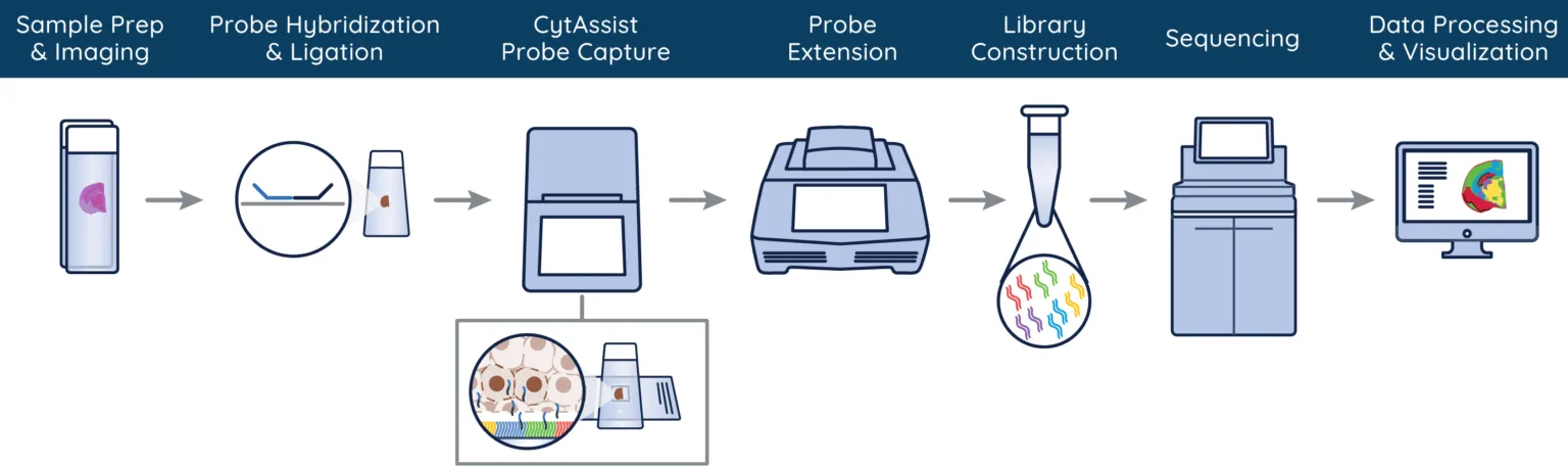
Figure 2. Visium CytAssist workflow from tissue sample processing to data analysis. Image from 10x Genomics.
Histopathology expertise provides several key benefits for spatial transcriptomics:
- Precise tissue selection: Pathologists can identify specific regions of interest within tissue samples, ensuring that spatial transcriptomics and proteomics captures relevant areas for gene expression analysis.
- Tissue handling expertise: Obtaining a high-quality stained image is crucial in the spatial transcriptomics workflow. AcelaBio has deep expertise in processing and embedding tissue, as well as sectioning and staining with established protocols to achieve best in class images.
- Tissue morphology assessment: Histopathology expertise allows for a detailed evaluation of tissue morphology, which is crucial for correlating spatial gene expression with structural and functional aspects of the tissue. This helps in understanding the spatial context of gene and protein expression patterns.
- Enhanced data interpretation: A pathologist’s ability to interpret staining techniques (e.g., H&E, immunofluorescence) provides valuable insights into how spatial gene expression correlates with protein expression and tissue architecture, enhancing the biological understanding of the data.
- Quality control: Histotechnologists can assess the quality of tissue sections and ensure that samples are appropriately prepared for spatial workflow, essential for obtaining high-quality reproducible results.
- Pathological relevance: In clinical studies, histopathological analysis can help identify disease-relevant regions, such as areas of inflammation or fibrosis, allowing for more targeted and meaningful spatial biology studies.
Incorporating spatial transcriptomics tools, such as the 10x Genomics Visium platform, into the drug development process enables a scientist to answer many questions.
- What cell types are present in a tissue and where are they located? Spatial transcriptomics allows scientists to identify different cell types within a tissue based on their unique gene expression profiles and helps determine the differentiation status of cells. This information can be mapped to specific locations within the tissue, providing a detailed atlas of cellular composition.
- Is a drug target present in a tissue where expected? Spatial transcriptomics can reveal which cellular pathways are altered in disease by analyzing and comparing gene expression patterns of healthy and diseased tissues. This information can help confirm presence and/or activation of a drug target for developing new therapies.
- Do histopathological changes correlate with expected gene expression changes? Spatial transcriptomics can correlate pathologist-identified features with changes in cell population composition and gene expression. This information is useful in determining how localized cell types can affect tissue morphology and disease progression.
- What is the difference in tissues of drug responders versus non-responders? Spatial transcriptomics can identify differences in cellular and molecular composition of patient tissues over the course of drug treatment, highlighting gene expression changes in cell populations over time. This information can provide insight into the response a patient has to a drug.
To find out more about spatial biology and Alimentiv/AcelaBio’s specialized services, visit our website or contact us.
References
- Marx, V. (2021) ‘Method of the year: Spatially resolved transcriptomics’, Nature Methods, 18(1), pp. 9–14. doi:10.1038/s41592-020-01033-y.
- Spatial Transcriptomics & Spatial Biology (no date) 10x Genomics. Available at: https://www.10xgenomics.com/spatial-transcriptomics (Accessed: 10 October 2024).